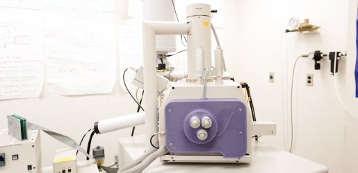
The FEI Philips XL 40 Environmental Scanning Microscope (ESEM) is a large-chamber, tungsten source, environmental scanning electron microscope capable of high and low vacuum imaging. The FEI Philips XL 40 ESEM is also capable of imaging hydrated and contaminated samples. Advanced accessories include a thin-window energy dispersive spectrometer (EDS) and hot or cold stages. The system is operated via easy-to-use software control using a Windows user interface. The ESEM can be used for organic and inorganic scanning electron analysis.
Capabilities
Remote Training and Collaboration
- New laboratory web camera
- Zoom screen-share abilities from both the microscope control and AzTEC analysis computers
- Remote technical assistance with Raritan DKX4-101 KVM-over-IP
- Remote operation with Raritan DKX4-101 KVM-over-IP
Instrumentation
- The NC/VP Materials Characterization Facility is home to the only environmental scanning electron microscope on campus.
- The instrument is capable of operating in low or high vacuum modes. It includes Oxford
AZtec EDS and both high (1,000 C) and low temperature stages.
- Remember to insert the EDS detector before use and remove it before venting. If the detector is not inserted, the counts will be VERY low.
- Insert the detector:
- On the AZtec computer, in the bottom right corner of the screen, click on the EDS icon and select Insertion then In.
- Remove the detector:
- On the AZtec computer, in the bottom right corner of the screen, click on the EDS icon and select Insertion then OUT.
- WARNING: This must be done before venting the chamber to prevent equipment damage.
- Large area montage imaging and elemental mapping (up to few square centimeters)
- EDS-based phase analysis
- Chemical contrast imaging and EDS-based particle analysis (Oxford AZtecFeature)
- Crystallographic phase mapping, texture analysis and grain size orientation distribution by Electron Backscatter Diffraction (EBSD, Oxford AZtecCrystal)
Training
Courses
Michigan Tech offers many undergraduate and graduate courses related to materials characterization.
Some of these courses offer direct, hands-on training in scanning electron microscopy.