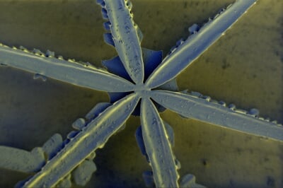
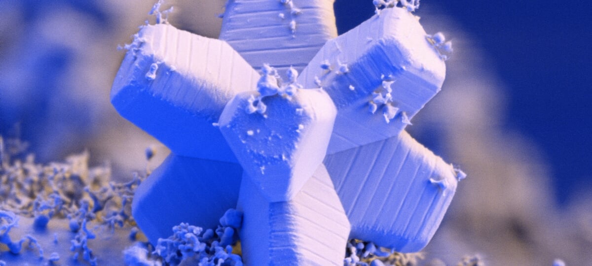
A unique laboratory at Michigan Tech captured microscopic photography of snowflakes in a demonstration of the lab's high-powered scanning electron microscope.
The Applied Chemical and Morphological Analysis Laboratory, known as ACMAL, is one of the most unique facilities at Michigan Technological University. When the lab's researchers went looking for a material that could demonstrate the capabilities of its scanning electron microscope, the natural choice was a one-of-a-kind resource that abundantly blankets campus every winter. The resulting snowflake imagery is an artful invitation to researchers on and off campus to discover the lab's resources.
Home to highly specialized and cutting-edge microanalytical and X-ray instruments, ACMAL is managed within Michigan Tech's Department of Materials Science and Engineering (MSE), but is designed to be shared by researchers from Tech and elsewhere.
ACMAL provides assistance and education for researchers interested in materials characterization through training and use of its shared instruments. The possibilities are nearly as limitless as the shapes of snowflakes, encompassing research projects in materials science, microbiology, biomedical engineering, environmental science and more.
"ACMAL is home to several very specialized cutting-edge pieces of equipment that are critical to leverage important research in many different fields," said ACMAL staff member Erico Freitas, an MSE research scientist and adjunct assistant professor. "ACMAL also offers several training programs to educate people on optical and electron microscopy, so faculty and students can learn how to operate several instruments independently."
The training benefit available to Michigan Tech students, faculty and staff also includes course support in numerous disciplines, including materials science, engineering, biomedical engineering, physics, chemical engineering, chemistry and biological sciences.
ACMAL's instrument inventory includes an atomic force microscope, a confocal microscope, flow cytometer, an X-ray photoelectron spectrometer, X-ray diffractometers, nano indentures, sample preparation equipment and several electron microscopes, including the one used recently to capture snowflake images.

As part of a cryo-microscopy training at ACMAL, Joey Tomei, MSE research scientist and ACMAL staff member, chose snowflakes to demonstrate the uses of the Thermo Fisher Apreo 2 field emission scanning electron microscope. It's an advanced scanning electron microscope designed to make high-performance imaging and analytics accessible to all users. It was a demonstration of accessibility in more ways than one. Tomei retrieved the demo material from right outside the Minerals and Materials Engineering Building, where many of ACMAL's instruments are located on the sixth floor.
During the session, Tomei showed the trainees how to transfer the snow in a small beaker kept cold to prevent any melting. In the microscope room, the snow was then plunged into liquid nitrogen to freeze the specimen to negative 175 degrees Celsius. It was kept at that temperature for the duration of the experiment. Held within a cryo preparation chamber, the scanning electron microscope was able to capture the intricate details of the snowflakes. The sample was later artificially colored using the scanning electron microscope's own imaging software. The images were shared on ACMAL's website, which, along with the training itself, provides outreach to encourage more researchers to make use of the lab's capabilities.
The scanning electron microscope, for example, can be used for much more than snowflake imaging. Combining super-high-resolution imaging capabilities with X-ray spectrometry analysis and electron diffraction, the microscope provides a comprehensive characterization of countless types of materials.
"Biological samples, polymers and other soft materials can be imaged and investigated under the scanning electron microscope," said Freitas. "It is an important characterization tool to investigate steel, metals and a variety of alloys. When compared to optical microscopy, the scanning electron microscope has a great advantage, as it allows imaging analysis with a much larger depth of field with much better spatial resolution."
The scanning electron microscope is not the only advantage ACMAL has at its disposal. Freitas manages the FEI 200-kilovolt Titan Themis scanning transmission electron microscope, or S-TEM — one of only two available at Michigan universities. This microscope, capable of imaging for structural and chemical analysis at the nanoscale, is housed in the Advanced Technology Development Complex, where the S-TEM has its own dedicated and stabilized space, complete with water-cooled temperature controls and backup power.

Under the Titan Lens
In addition to high-resolution images, the S-TEM can also perform fractional or chemical analysis. Its applications are useful in many research areas, like biomedical engineering, materials science, and chemical sciences.
Having access to both advanced instruments and trained specialists who manage them and can provide training gives Michigan Tech researchers valuable opportunities to further their work.
"I could spend several years on a daily basis working on these microscopes and still not be able to get all they may offer in terms of characterization possibilities," said Freitas. "I love the fact that I can help educate people on electron microscopy, training people or teaching the electron microscopy classes."
Michigan Technological University is an R1 public research university founded in 1885 in Houghton, and is home to nearly 7,500 students from more than 60 countries around the world. Consistently ranked among the best universities in the country for return on investment, Michigan's flagship technological university offers more than 120 undergraduate and graduate degree programs in science and technology, engineering, computing, forestry, business, health professions, humanities, mathematics, social sciences, and the arts. The rural campus is situated just miles from Lake Superior in Michigan's Upper Peninsula, offering year-round opportunities for outdoor adventure.


Comments