FE-SEM
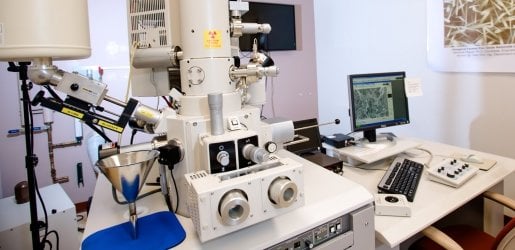
The Hitachi S-4700 FE-SEM is a cold field emission high resolution scanning electron microscope.
The Hitachi S-4700 FE-SEM is a cold field emission high resolution scanning electron microscope. This SEM permits ultra high resolution imaging of thin films and semi-conductor materials on exceptionally clean specimens. It is also suitable for polymeric materials. The S-4700 is configured to detect secondary and backscattered electrons as well as characteristic X-rays. The system is fully automated and is operated via easy-to-use menu driven software.
There is a new Linux computer in the FESEM. Below are instructions for data transfer from the FESEM:
Note: Only use the provided USB for data transfer from the FESEM.
Use the software only when you do the alignments. Do not change the physical knobs of the apertures.
The beam monitor aperture is set at position 2 and should be kept at 2.
If you ever need to change the objective aperture, only use the knob to switch between apertures 1, 3, or 4. Do not use aperture 2.
Objective aperture sizes: 1 > 3 = 2 > 4
Attn.: Objective apertures 2 and 3 have the same size. We exchange them annually when one or the other gets dirty. This year (2024–25) we are using 3.
Home stage position is:
4pi Analysis, Inc. engineers and sells X-ray Microanalysis and Digital Imaging Systems. (NOTE: 4pi has ceased manufacturing and sales. ACMAL is confident that this will not affect 4pi’s ability to provide technical support to this lab.)
Free online eTraining is available for this instrument. This self-paced tutorial and reference content does not replace course requirements for authorized usage.
Hitachi S-4700 FE-SEM Online Training
Michigan Tech offers many undergraduate and graduate courses related to materials characterization.
Current concepts in light and electron microscopy and scanning probe techniques. Theory and practice of fluorescence (including confocal and multi-photon), atomic force, scanning and transmission electron, and video microscopy as applied to biological specimens with emphasis on sample preparation. Half semester course.
Current concepts in light and electron microscopy and scanning probe techniques. Theory and practice of fluorescence (including confocal and multi-photon), atomic force, scanning and transmission electron, and video microscopy as applied to biological specimens with an emphasis on sample preparation. Emphasis will be placed on application of advanced techniques. Half semester course.
A study of recent developments in the biological sciences.
A discussion of recent developments in the biological sciences. Recent offerings have included population genetics, taxonomy of aquatic insects, herpetology, bryology, fungi, and lichens.