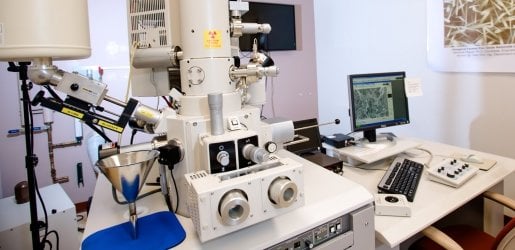
Electron Gun
The gun is composed of a cold field emission tip and two anodes. It generates electrons and accelerates them to an energy in the range of 0.1–30 keV (100–30,000 electron volts).
Beam Monitor Aperture
This aperture is a passive current monitor that measures the probe current. It is used to detect current fluctuations, which are used as a reference signal to stabilize or reduce noise in that image.
Anti-Contamination Trap Dewar
Also called a cold trap, it prevents specimen contamination by hydrocarbon build-up during high magnification, or low accelerating voltage, specimen viewing. The trap is a plate above the specimen that adsorbs gas around the specimen.
Upper Secondary Electron Detector
Also known as Everhart-Thornley detector. The upper detector is located above the objective lens and attracts high-resolution secondary electrons. Its function is to collect secondary electrons from the specimen surface, view images from above to easily see areas of low topography, and to give clean images using its high signal to noise ratio.
Objective Aperture Assembly
Composed of the aperture selector and aperture translation knobs, it allows for the selection of aperture size and alignment of the electron beam through the objective lens.
Liquid Nitrogen Tank
Contains the liquid nitrogen for cooling the EDS detector and reducing the thermally induced background, or "leakage" current, present in the signal. A cold finger and cold trap are also used to keep the environment clean.
Gun Valve
A pneumatic valve that automatically closes in the event of accidental specimen chamber venting. This ensures that the environment of the electron gun remains uncontaminated.
EDS Detector
The energy-dispersive spectrometer is a semi-conductor based detector that measures X-ray signals. It can simultaneously detect characteristic X-rays of all elements above atomic number 4 at higher beam currents than used for secondary electron imaging.
Lower Secondary Electron Detector
Also known as Everhart-Thornley detector. It is located inside the specimen chamber, attracting largely backscattered electrons that create a topographic signal. It collects secondary electrons from the specimen surface as well as the surrounding area. It provides a side view of the specimen, showing irregular surfaces.